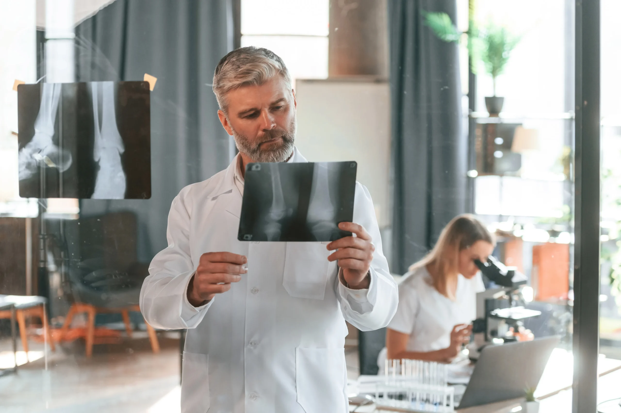
In the last decade, 3D MRI imaging has emerged as one of the most transformative tools in the realm of medical visualization. By converting traditional 2D MRI slices into comprehensive 3D body scans, clinicians and specialists can now gain a holistic understanding of patient anatomy with remarkable precision.
Understanding the Basics of MRI and CT Imaging
MRI (Magnetic Resonance Imaging) and CT (Computed Tomography) imaging have long been staples of modern medicine. CT MRI imaging allows healthcare providers to peer inside the human body non-invasively, providing critical data for treatment planning and monitoring. What separates traditional scans from 3D medical imaging is the capacity for depth, rotation, and spatial orientation.
The 3D Advantage
With the integration of 3D MRI and CT technologies, medical imaging software has taken a massive leap forward. By using advanced rendering algorithms, 3D medical imaging software turns standard image data into rotatable, zoomable models that show bones, soft tissue, and structural relationships with startling clarity. This leap has enabled professionals to engage more meaningfully with the images—exploring anatomy from angles that were once limited to imagination.
Applications Across Fields
From sports injury recovery to pre-operative planning, the use of 3D medical imaging is expanding fast. It’s also a game-changer in education, where students can interact with detailed, accurate models without needing access to live cases.
Enhancing Communication
Perhaps one of the most underrated benefits of 3D body scan technology is its ability to improve communication. Whether it’s between medical professionals or from specialist to patient, being able to visually demonstrate anatomical features creates shared understanding.
Innovations in Medical Imaging Software
Today’s top-tier 3D medical imaging software is browser-based, secure, and lightning-fast. Some platforms now allow for side-by-side comparisons of different scans, time-lapse imaging, and detailed annotation tools—all of which contribute to better record-keeping and collaboration.
